
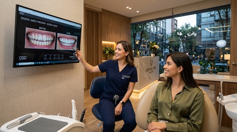
At Clínica DrDiente Polanco, dental 3D scanning is used to create a detailed digital record of teeth, gums, and bite without traditional impression material. Located in Polanco, CDMX, the clinic uses this information as part of an educational and diagnostic process for treatments such as smile design, implants, and invisible orthodontics. This article explains what a scan can show, what it cannot show on its own, and why the clinical evaluation still matters.
The perspective of Dr. Carlos Ariza on dental 3D scanning
Dr. Carlos Ariza’s clinical approach at Clínica DrDiente is based on planning before treatment. In simple terms, an intraoral scan is like making a highly detailed digital map before choosing a route: it helps the dental team see the terrain, compare options, and communicate the proposed plan more clearly.
The short answer is that scanning can make treatment planning more visual and more comfortable for many patients. An iTero intraoral scanner captures images of the teeth, gums, and bite to build a three-dimensional model. This model may support planning for digital smile design, crowns, aligners, and selected implant cases.
What matters is not simply having advanced technology. The value comes from how the information is interpreted alongside an examination, photographs, bite assessment, and any radiographs that are clinically justified. There is no single answer for everyone. A scan is a tool; it is not a diagnosis by itself.
Here is something many patients do not hear at the first appointment: a beautiful digital simulation should never be treated as a promise of a final result. It is a planning conversation. Later in this article, that distinction becomes especially relevant for veneers and implant planning.
What most dentists do not tell you about dental 3D scanning
Dental 3D scanning does not use radiation. A dental CT scan, also called CBCT or cone-beam computed tomography, does use X-rays. They are different studies with different purposes. A CBCT should not be added automatically just because an intraoral scan is being performed; it should be requested when it can change a clinical decision, such as evaluating bone volume or anatomical structures before an implant procedure.
For implant planning, the optical scan may be combined with a justified CBCT study. This can help visualize the tooth surfaces together with bone, roots, the maxillary sinus, and the mandibular nerve canal. Guided surgery can then be planned with a surgical guide. Still, guided planning has limits. A 2025 clinical trial reported mean deviations in guided implant placement, so the guide does not replace professional judgment, safety margins, or follow-up care. Read the clinical trial on guided implant surgery.
For crowns, a 2024 randomized trial found better average marginal fit for posterior crowns made from intraoral scans than from conventional impressions followed by model digitization. That evidence applies to the type of restoration studied; it is not a guarantee for every mouth or every rehabilitation. Review the posterior crown clinical trial.
What nobody tells you is that the scan is only as useful as the conditions during capture. Saliva, bleeding, deep margins, tooth movement, missing teeth, and severe grinding can make records more difficult. In practice, the team may repeat an area, use complementary techniques, or recommend a conventional impression when that produces a more reliable record.
The open question from earlier has an answer now: a simulation is useful because it helps people understand possibilities, but it cannot replace healthy gums, stable bite function, or careful treatment sequencing.
Our clinical experience: hypothetical examples
The following examples are hypothetical educational scenarios, not documented Clínica DrDiente cases or patient testimonials. They illustrate how digital records may guide conversations in a Polanco dental consultation.
A hypothetical 34-year-old patient asks for veneers because her front teeth look uneven in photos. The scan and clinical photographs may help compare tooth proportions and plan a temporary mock-up. If the gums are inflamed, have plaque accumulation, or are not a healthy coral-pink color, veneer treatment should wait. The same applies when there are cavities or an unstable bite. First, the mouth needs to be treated and stabilized.
A second hypothetical patient wants teeth that are very white and perfectly square. Digital planning can be helpful for showing why natural teeth are not flat squares or “Adams chewing gums.” Natural-looking teeth have curves, textures, and variation. A good smile plan should look like it belongs to the person, not like a uniform artificial template.
A third hypothetical patient is considering an implant after losing a tooth. A scan may record the neighboring teeth and bite, while a clinically indicated CBCT may assess bone and nearby anatomy. The proposed plan can then be reviewed before any irreversible decision. Patients can also explore documented examples in the DrDiente real-case gallery.
For ceramic veneers, digital planning can help define preparation. When preparation is appropriate, it should be conservative and individualized. Clínica DrDiente describes minimally invasive, ultra-precise preparation calibrated between 0.3 and 0.5 mm in its own laboratory workflow. Even then, this is not automatically suitable for every patient.
Frequently asked questions about dental 3D scanning
Is it normal to feel nervous about an intraoral scan?
Yes. Many people expect something similar to a traditional impression tray. In simple terms, the scanner is moved around the teeth to capture images. It can be more comfortable for patients who experience gagging with molds, although comfort varies from person to person.
Does a dental 3D scan hurt?
The short answer is no: intraoral scanning is non-invasive and does not use radiation. If gums are tender or inflamed, the area may feel sensitive during any dental examination. The clinician should adapt the appointment to the patient’s comfort.
How long does a dental 3D scan take?
It depends on the area being scanned, saliva control, the bite record, and whether records need to be repeated. A single scan may be relatively quick, while full planning with photographs, discussion, and additional clinically justified imaging takes longer.
What happens if the scan shows something unusual?
A scan may reveal shape, alignment, wear patterns, and bite relationships, but it does not independently detect every cavity, pulp infection, or bone-quality issue. The next step depends on the clinical examination and, when justified, appropriate radiographic imaging.
How do I know whether I need a CT scan too?
For an implant, complex surgery, or another situation where bone anatomy may change the plan, a CBCT may be indicated. For routine alignment planning or a crown, it may not be necessary. Ask what decision the CT scan will help make and whether the smallest appropriate field of view will be used.
Where can I receive dental 3D scanning in Polanco?
Clínica DrDiente Polanco is located at Av. Presidente Masaryk 83, Polanco V Secc., Miguel Hidalgo, CDMX. Patients should confirm the type of evaluation needed, including whether a CBCT is clinically indicated, before their visit.
What technology does DrDiente use for dental 3D scanning?
Clínica DrDiente Polanco describes a digital protocol that may include iTero intraoral scanning, clinical photography, virtual design and wax-up planning, and CAD/CAM CEREC technology. For selected cases, tomographic 3D imaging may be used when clinically indicated. These records can support planning for crowns, restorations, invisible aligners, smile design, and implants.
The key is in combining information rather than relying on one screen. An intraoral scan shows the visible surfaces of teeth and gums. Photographs show facial and smile context. A bite evaluation helps assess function. A CBCT, when indicated, contributes information about bone and internal anatomy. This is particularly relevant for dental implant planning and may also help patients considering invisible aligners.
Patients visiting CDMX from abroad for dental tourism should approach digital dentistry with the same questions as local patients: What is the diagnosis? Which records are necessary? What alternatives preserve more tooth structure? Can I receive copies of my images and treatment plan? For information about this route, visit dental tourism in CDMX.
Dental technology can improve communication and planning, but it does not make care automatic. Healthy gums, caries control, bite stability, and a realistic treatment goal remain the basis of responsible dentistry.
Schedule with the DrDiente team
If you want a second opinion or would like to begin a treatment conversation, Clínica DrDiente Polanco can explain how digital records may fit your specific situation. The consultation is educational and does not replace an individualized clinical diagnosis.

Information from Clínica DrDiente
Aesthetic Dentistry & Oral Rehabilitation · COFEPRIS 2409132002A00145
This content is informational and is not a substitute for professional dental advice. Book an assessment for a personalized diagnosis.
