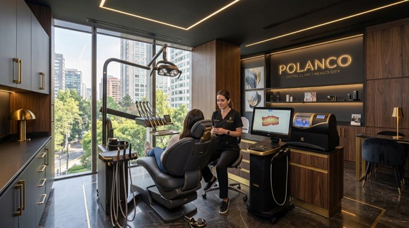
At Clínica DrDiente Polanco, patients often ask whether an intraoral scanner is better than traditional dental impressions. The short answer is: it depends on the treatment, the condition of the gums, and what needs to be made. A digital scan can be remarkably comfortable and useful, but it is not a replacement for a clinical examination, X-rays, or periodontal evaluation. Dr. Carlos Ariza and the team use digital planning as part of a careful approach to dentistry in Polanco, CDMX.
In simple terms, a traditional impression uses a tray and a soft material that records the teeth and gums. That record is usually poured in plaster or digitized later. An intraoral scanner projects light, captures thousands of images, and creates a three-dimensional digital model of the mouth. Both methods are ways to collect information. Neither one, by itself, is a treatment.
The perspective of Dr. Carlos Ariza on intraoral scanning and dental impressions
A good dental record is not about choosing the newest-looking tool. What matters is whether the information is reliable enough for the planned restoration, aligner, crown, implant, or smile design. At Clínica DrDiente, digital tools such as an intraoral scanner, photographic protocol, and 3D imaging can help organize that information more clearly. Still, technology only works well when the mouth is ready to be scanned.
Is it normal to prefer a scan because impression material causes nausea? Yes. Many people find the scanner more comfortable because there is no tray filled with material covering the palate. If a small area is missing, it can often be captured again without repeating the entire record. That can make a real difference for someone with a strong gag reflex.
There is something many patients are not told at the first appointment: a beautiful digital model can still be incomplete if the gums are bleeding, saliva is difficult to control, or a restoration edge is hidden under inflamed tissue. The scanner sees what it can see. It cannot guess what is covered by blood, plaque, or swollen gum tissue. Later in this article, that point becomes especially relevant for veneers and crowns.
What most dentists do not tell you about intraoral scanners and traditional impressions
Digital does not automatically mean more accurate in every situation. For a single crown or a short bridge, both methods can provide clinically acceptable results when performed correctly. A randomized clinical study comparing posterior crowns found a better average marginal fit with intraoral scanning than with conventional impressions followed by scanning the plaster model: 57.94 ± 22.51 micrometers versus 82.98 ± 21.72 micrometers. The digital workflow also required fewer clinical adjustments in that study. Read the clinical study.
That does not mean every patient should receive a scan only. What happens if someone needs several implants, a full arch restoration, or a complete denture? The answer changes. With multiple implants, the distance between implants and the verification method can affect precision. For complete dentures, mobile soft tissue may need a functional impression, meaning a conventional material can record how the tissue moves under normal conditions. In those cases, a hybrid workflow may be more sensible.
Think of it like tailoring a suit. A digital measurement can be excellent, but if the person moves, the fabric shifts, or the body position is not represented properly, the final fit still needs checking. In dentistry, the fit is not judged by a screen alone. It is checked clinically, at the tooth or implant, before a restoration is finalized.
For implant-supported restorations, a clinical trial found no statistically significant difference in positional deviation between intraoral scanning and open-tray impressions for three-unit prostheses. Both methods were considered acceptable in that setting. See the implant clinical trial. This is why there is no single answer for everyone.
The open question from above has a practical answer: healthy gums are part of scan accuracy. Before veneers or a digital smile design, there should be no active cavities, heavy plaque, or inflamed gums. Healthy gums should look coral pink, not swollen or prone to bleeding. If gum tissue is inflamed, placing veneers is not an ethical shortcut. It is like having a painful hangnail: the tissue stays irritated because something is invading or disturbing its space.
Our clinical experience: real-case considerations
To be transparent, the following examples are hypothetical educational scenarios, not real patient cases or testimonials. They illustrate why the same device may be used differently from one person to another.
A hypothetical 34-year-old patient needs one posterior crown and has healthy gums, good access, and no heavy saliva flow. An intraoral scan may allow the clinician and laboratory to review the preparation immediately and manufacture the crown through a digital workflow. The final result still requires an in-mouth fit check.
A hypothetical patient seeking invisible aligners may benefit from a digital scan because the three-dimensional model can support treatment planning and progress records. How long does the scan take? Often only a few minutes of active scanning, although the total appointment depends on photographs, bite records, examination, and treatment planning.
A hypothetical patient missing many teeth may need a more complex approach. A scan can be valuable for diagnosis and primary records, while conventional impressions, verification steps, or other techniques may still be needed before a full-arch implant restoration is made. In complete-arch cases, precision becomes more demanding as the span increases.
Patients can view documented visual outcomes only through the Clínica DrDiente gallery of real cases. A photograph alone does not reveal the full diagnosis, gum health, bite relationship, or maintenance needed for each person.
Questions I receive frequently about intraoral scans and impressions
Is an intraoral scan painful?
Usually, no. The scanner tip moves around the teeth and does not use needles or impression paste. Some people may feel temporary discomfort if they have sensitive teeth, limited opening, or sore gums. For an honest answer, the level of comfort depends on the condition of the mouth and the person’s gag reflex.
How do I know if I need a scanner instead of a traditional impression?
The short answer is that you do not need to choose before the evaluation. The planned treatment guides the decision. Scans are commonly useful for crowns, veneers, short restorations, aligners, retainers, and digital smile planning. Traditional impressions may remain useful when capturing mobile gum tissue, certain complete dentures, or complex implant cases.
What happens if my gums bleed during the scan?
Bleeding can block the area that needs to be recorded. The clinician may pause, improve gum health, control moisture, or use a different recording method. Technology should not be used to hide a biological problem. The key is to treat the cause before proceeding with a definitive restoration.
Is a digital scan worth it for veneers?
It can be, especially when combined with photographs and digital planning. But a scan does not make veneers appropriate by itself. Veneers should not be placed over active decay, inflamed gums, or an unstable bite. If alignment or malocclusion is the issue, orthodontic treatment may come first.
Where can I be treated for an intraoral scan in Polanco, CDMX?
Patients looking for a dentist in the area can schedule an evaluation at Clínica DrDiente Polanco, Av. Presidente Masaryk 83, Polanco V Secc., Miguel Hidalgo, CDMX. The appointment should include a clinical assessment, not just a digital scan, so the recommended method matches the treatment goal.
Now the earlier point is complete: the scanner is only as useful as the preparation around it. A clear field, stable tissues, and a defined treatment plan matter more than simply choosing “digital.”
What technology do we use at DrDiente for intraoral scanning and impressions?
At Clínica DrDiente, digital diagnosis can combine an intraoral scan with a photographic protocol and, when clinically indicated, 3D tomography. Tomography is a three-dimensional X-ray that can help evaluate bone and anatomical structures for implant planning. It is not necessary for every scan or every treatment.
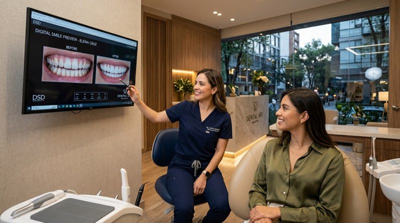
For smile design, the digital record helps evaluate tooth proportions, bite, facial features, and planned changes before any irreversible work is considered. Natural-looking teeth are not square or artificial like “Adams chewing gum.” They have curves, texture, and individual character. When tooth preparation is indicated for veneers, it should be conservative and measured; planning may target approximately 0.3 to 0.5 mm of enamel preparation, depending on the case and planned material.
That precision does not authorize unnecessary treatment. A scan supports decisions; it does not make them for the clinician or patient. For international patients researching dental tourism CDMX, it can also make it easier to organize records with a laboratory or treating dentist, while protecting clinical information in the patient record. Mexico’s NOM-004-SSA3-2012 recognizes electronic clinical records and requires their appropriate handling and confidentiality. Review the official standard.
Agenda with the DrDiente team in Polanco
If you want a second opinion or want to begin treatment, schedule an evaluation with the Clínica DrDiente team in Polanco, CDMX. The goal is not to sell a scan or an impression; it is to determine which record will support a safe, precise plan for your mouth. All open questions in this guide come back to the same idea: the right method depends on the diagnosis, healthy tissues, and the restoration being planned.

Information from Clínica DrDiente
Aesthetic Dentistry & Oral Rehabilitation · COFEPRIS 2409132002A00145
This content is informational and is not a substitute for professional dental advice. Book an assessment for a personalized diagnosis.