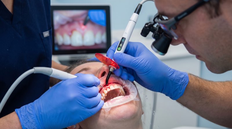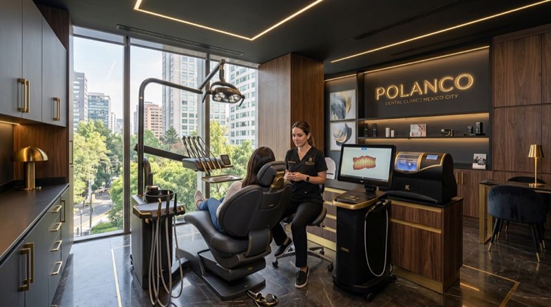
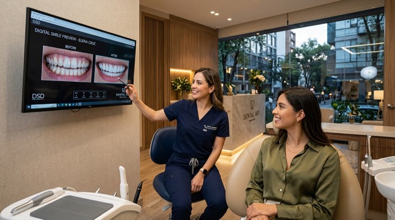
Yes. Clínica DrDiente Polanco uses diagnostic dental photography as part of treatment assessment and planning. Professional images can help document the relationship between the smile, lips, face, gums, tooth color, and dental proportions. At the Polanco, CDMX location, photography may be combined with an intraoral scan, digital planning, and 3D tomography when there is a clinical reason to use it. This is educational information, not a diagnosis.
Dr. Carlos Ariza’s perspective on dental photography for treatment planning
Dr. Carlos Ariza is associated with Clínica DrDiente’s approach to digital diagnosis: treatment decisions should begin with information, not assumptions. In simple terms, a professional dental photo is like looking at a room with the lights on instead of trying to arrange furniture in the dark. It can show details that are difficult to judge from memory, a quick mirror check, or an unstandardized cellphone image.
The short answer is that photography can improve communication and planning, but it does not replace an examination. A photo cannot measure every periodontal detail, identify all cavities, assess bone volume, or explain how the bite functions. What matters is how the image fits with the patient’s oral examination, medical history, bite assessment, radiographs, and, when justified, cone beam 3D tomography.
At Clínica DrDiente Polanco, diagnostic photographs are described as part of the initial assessment. They can be used to evaluate facial and smile proportions, gum display, tooth shape, texture, and shade before discussing options such as digital smile design and veneers, crowns, aligners, or implant rehabilitation. There is one detail many patients do not hear at the first appointment: a beautiful simulation is not a guarantee of a clinical result. Gum biology, tooth structure, bite forces, healing, and laboratory work still influence the final outcome.
What most dentists do not tell you about dental photography
Professional dental photography is not simply taking flattering photos of teeth. A useful protocol normally includes extraoral images of the face and smile, plus intraoral images using mirrors and retractors. These images can help identify a shifted dental midline, uneven gum display, old restorations, spacing, wear from grinding, fractures, color differences, and the relationship between the teeth and lips.
“Is it normal for my smile to look different in photos?” Yes, sometimes. Lighting, camera angle, lip position, and even the way a person smiles can change what is visible. That is why standardized photography matters. White balance, lighting, magnification, polarization, and calibration can affect how tooth color appears. A cellphone photo may be useful for communication, but it is not automatically equivalent to professional clinical photography.
For honest planning, photography must be paired with direct examination. If a person has gum inflammation, cavities, plaque accumulation, or an unstable bite, cosmetic work should wait. Healthy gums should look coral pink rather than red, swollen, or prone to bleeding. Placing veneers over inflamed gums is like ignoring a painful hangnail: the irritation does not disappear because something attractive covers it. It can continue to swell and bleed.
“How do I know if I need dental photography?” It depends on the treatment. It is especially helpful before a smile design, veneers, crowns in the front teeth, orthodontic planning, complex rehabilitation, or implant treatment in an aesthetic area. For implants, photographs help plan the visible smile component, while bone quantity and three-dimensional implant position require imaging such as CBCT only when clinically indicated. The loop closes here: photography supports a better conversation, but it never substitutes for the biological and functional assessment behind that conversation.
Our clinical experience: hypothetical examples
To avoid presenting undocumented cases as real patient stories, the following examples are hypothetical. They illustrate situations that may be discussed during an evaluation at Clínica DrDiente Polanco.
Hypothetical example 1: A 34-year-old patient asks for veneers because the upper front teeth look uneven in selfies. Standardized photos and an intraoral scan could show that the main issue is tooth position and a narrow smile rather than tooth color alone. In that situation, invisible aligners may be discussed before any restorative option. No single answer works for everyone.
Hypothetical example 2: A patient has a darkened front tooth and an older restoration with a visible edge. Photographs may help compare shape, translucency, texture, and color with neighboring teeth. A shade guide and other validated color tools may still be needed. The key is to avoid creating teeth that look flat, overly white, or artificial.
Hypothetical example 3: A patient with a missing tooth wants an implant immediately. Facial and smile photographs can help plan the visible result, while tomography may be necessary to answer a different question: whether bone dimensions and anatomy support the proposed implant position. This is why a photograph alone cannot approve or rule out implant treatment.
For documented outcomes, patients can review the DrDiente gallery of real cases and ask what records, diagnosis, and follow-up information apply to each treatment category.
Frequently asked questions about dental photography
Does professional dental photography hurt?
No. The short answer is no. Mirrors, retractors, and positioning may feel unfamiliar, and some people experience mild temporary dryness around the lips, but photography itself does not involve drilling or injections.
How long does dental photography take?
It depends on the type of records needed. A focused photo series may take only a short part of an appointment, while a complete diagnostic workflow can take longer because it may include an intraoral scan, clinical evaluation, bite records, and imaging when appropriate.
What happens if the photos show gum inflammation or cavities?
Treatment planning should address health first. Cleaning, gum care, cavity treatment, or bite stabilization may be recommended before cosmetic dentistry. In practice, starting veneers or a smile design before the mouth is stable can create avoidable complications.
Are photos enough to choose veneers?
No. Photography can help visualize proportions and expectations, but it cannot determine veneer suitability by itself. Available enamel, gum health, tooth alignment, cavities, grinding habits, and bite relationship all need assessment. Teeth should not look square like “Adams gum”; natural teeth have curves, texture, translucency, and small individual differences.
Is it worth getting a mock-up before changing my smile?
For many patients, it may be worth discussing. A mock-up is a reversible preview that can help assess form and length before irreversible preparation. If veneers are clinically appropriate, preparation should be conservative and individualized. Clínica DrDiente describes minimally invasive, ultra-precise preparation calibrated from 0.3 to 0.5 mm in its in-house laboratory workflow; suitability still depends on the individual tooth and treatment plan.
Where can I receive treatment planning with dental photography in Polanco?
Patients can request an evaluation at Clínica DrDiente Polanco, located at Av. Presidente Masaryk 83, Polanco V Secc., Miguel Hidalgo, CDMX. Ask which records are recommended for your specific concern, why each image is needed, and whether you can receive a written plan with alternatives, risks, maintenance needs, and expected limitations.
What technology does DrDiente use for dental photography planning?
Clínica DrDiente describes a digital diagnostic workflow that may include professional photographic records, intraoral scanning, virtual design or waxing, and 3D tomography when clinically justified. The intraoral scan creates a digital model of the teeth. Tomography produces three-dimensional imaging that can assist with questions involving bone, roots, and anatomical structures. A photographic protocol adds the visible facial and smile information that a scan alone cannot provide.
For a smile design, this combination can help the team communicate with the dental laboratory and with the patient. Technology of this kind is useful because it makes planning more visible and reproducible; it does not make every treatment automatic. A 2024 randomized clinical trial reported higher average satisfaction and more excellent ratings for a digital workflow than a conventional workflow in that study population, but one study does not mean digital treatment is superior for every patient. Read the clinical trial on PubMed.
For color matching, professional images should be interpreted carefully. Lighting and calibration affect the result, so photographs may be combined with a shade guide and other validated methods where available. A recent in-vitro comparison found improved color consistency with standardized camera and phone approaches compared with visual selection, though clinical confirmation in patients is still needed. Read the 2026 color comparison study.
Clínica DrDiente also provides care connected with smile design, implants, and invisible orthodontics, including planning for patients interested in dental tourism in CDMX. The practical point is simple: advanced technology should answer a clinical question, not be used because it looks impressive. That is the final piece of the earlier open loop. Images help reveal possibilities, while a responsible plan respects function, biology, and limits.
Treatment planning in Polanco, CDMX
If you are considering cosmetic dentistry, orthodontics, implants, or a second opinion in Polanco, ask whether your assessment includes standardized photographs and how those images will influence the plan. You may also ask whether gums are healthy, whether a mock-up is appropriate, whether tooth movement should come before restorations, and what parts of the proposed treatment are irreversible.
In Mexico, photographs may form part of the clinical record. Identifiable images should be handled confidentially, and written authorization is needed if photographs are published or otherwise disclosed. The applicable medical-record standard also addresses confidentiality and retention requirements. Consult NOM-004-SSA3-2012.
Schedule with the DrDiente team
If you want a second opinion or would like to begin a treatment evaluation, contact Clínica DrDiente Polanco. A consultation can clarify whether professional photography, digital scanning, tomography, or another record is appropriate for your goals and oral health. Visit the Polanco location at Av. Presidente Masaryk 83, Polanco V Secc., Miguel Hidalgo, CDMX.

Information from Clínica DrDiente
Aesthetic Dentistry & Oral Rehabilitation · COFEPRIS 2409132002A00145
This content is informational and is not a substitute for professional dental advice. Book an assessment for a personalized diagnosis.